
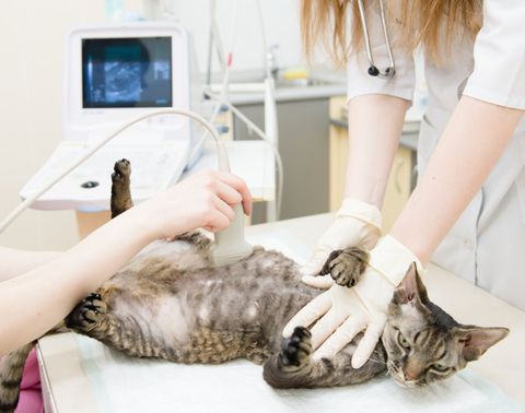
Ultrasound
Ultrasound scanning or sonography, involves the use of sound waves to produce pictures of the inside of the body. We often use this type of imaging when looking at the abdomen because ultrasound images are captured in real-time. They can show the structure of the body's internal organs and may provide key diagnostic clues in trying to obtain a correct diagnosis. Organs we commonly look at with ultrasound include: the kidneys, spleen, liver, gallbladder, pancreas, stomach, intestines, bladder, as well as blood flowing through blood vessels. Ultrasound imaging can be performed using minimal restraint or sedation.
Echocardiogram
Echocardiogram or commonly referred to as “echo” is ultrasonography specific to the heart. It allows us to watch the movement of how the parts of the heart work and where the blood is going. Echocardiography is the minimally invasive gold standard to identify the source of most cardiac abnormalities. This imaging is not painful, noninvasive, and are performed while your pet remains awake. These crucial diagnostics offer the cardiologist a great deal of information regarding the structure and function of the heart. Once the condition is properly diagnosed, treatment can begin.